Common Hip Conditions
Degenerative Osteoarthritis (OA)
Degenerative osteoarthritis (OA) is the most common disease process resulting in the need for a Total Hip Replacement (THR). Degenerative Osteoarthritis is a progressive joint disease that affects people’s ability to move their hip joint without pain. While OA is more common as we age, not everyone develops OA.There are two types: primary and secondary. While the symptoms are the same for the both types, the underlying cause is different.
Secondary Degenerative OA
Secondary Degenerative OA is the result of a prior trauma or an underlying condition that altered the hip’s normal architecture, resulting in joint deterioration and arthritis. Often a patient has subtle congenital or early childhood abnormalities which alter normal hip biomechanics and result in a slow progressive destruction. The person may not have even been aware of the condition until the hip became symptomatic many decades later.
In our practice, the majority of hips with degenerative OA are secondary to an underlying condition. We commonly see patients who had developmental conditions at birth, such as hip dislocation or subluxation, or who developed conditions during childhood when their skeletons were still maturing.
Some people are born with subtle abnormalities in the hip socket area that cause the femoral head or femoral neck to “bump” into the rim of the socket when they move. This can damage the rim and lead to arthritis. This condition is referred to as acetabular femoral impingement (FAI) and is a common cause of secondary OA. (will link to blog on subject)
Prior hip trauma with or without a fracture, failed prior surgery on the hip, autoimmune inflammatory conditions like rheumatoid or psoriatic arthritis, loss of the blood supply to the femoral head as an adult (AVN or osteonecrosis) or as a child (Legg-Perthes disease) or a slipped capital femoral epiphysis all are common conditions leading to a diagnosis of secondary OA and the need for a total hip replacement (THR) later in life.
Primary Degenerative OA
Primary Degenerative OA, a less common condition, occurs when hips develop degenerative arthritis without a clear underlying cause. A genetic basis is often assumed, as many times patients report other members of their family developing OA and needing THR.
Failure of Prior Implant
Dr. Leone is often called on to operate on joints that have been operated on multiple times before by other surgeons. In all cases where revision is indicated, we work to understand why the implant failed and how to correct it. We undertake extensive preoperative planning, optimize the patient’s medical condition before, during and after surgery, surgically attempt to correct all the problems that led to the failure and finally re-build a stable mechanical construct that re-creates hip mechanics.
Common causes for failure include loosening, wear or breakage of the components, peri-prosthetic bone loss (osteolysis), infection, recurrent dislocation, leg length inequality, pain, corrosion, metal toxicity or fracture.
Dislocation
The prosthetic ball in the socket can dislocate because the components wear down, break, or become unstable due to suboptimal positioning in the skeleton and/or relative to each other. The good news is that advances in design and materials are leading to more stable, durable components. For example, the femoral head in the newer implants is typically larger and thus more stable, than it was just a few years ago. Also, the quality of the plastic bearing surface has improved, making it much more durable and resistant to wear. Alternate bearing surfaces have also been developed including those made of ceramics and metal. More refined surgical approaches and advances in soft tissue closure have also markedly lessened the chance of a dislocation after surgery.
Leg Length Inequality
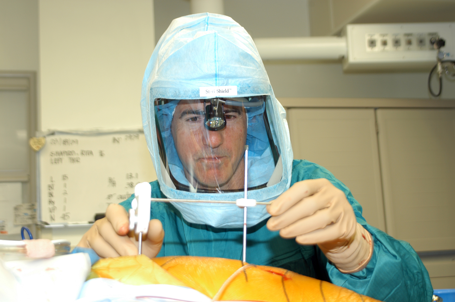
Creating equal leg lengths after surgery is a priority after THR and very much an “art”. If a patient’s legs are not equal in length after surgery due to improper implant positioning, instability and pain can result. Critical to reconstructing a stable hip with equal leg lengths is implanting the cup and stem in an optimal position in the bone and optimizing the length and tension in the surrounding soft tissues. At the Leone Center, we use the pelvic alignment level (PAL) to optimize positioning and leg length. This device, which Dr. Leone invented, has been a critical success factor in his ability to achieve consistent outcomes.
Osteolysis (Bone Loss)
Osteolysis is a condition in which the bone that supports the prosthesis is slowly destroyed, which causes the prosthesis to loosen. After millions of cycles and years of use, the prosthetic ball can wear down the liner of the socket, causing the release of tiny particles of metal or plastic debris. The “wear” debris accumulates in the tissue and causes inflammation which, in turn, stimulates bone cells to reabsorb bone, which in effect, destroys the bone supporting the prosthesis.
Better quality plastics and alternate bearing surfaces are reducing the incidence of osteolysis. Most modern prostheses are made of highly cross-linked polyethylene which is also helping to decrease the incidence and severity of osteolysis and resulting in improved prosthetic longevity.
Metal on Metal Implants
THR implants include a ball and socket. Various materials and combinations have been used over the years to produce these components, among them: balls made out of various metals and ceramics and cup liners made out of metals, plastics and ceramics. Today, most consider a ceramic ball articulating against a modern “highly cross linked polyethylene” socket to be the gold standard combination.
Many patients who have had “metal on metal” implants, that is a metal ball against a metal liner or socket, are now experiencing problems that require revision. We are also seeing issues with patients who have a hip resurfacing. Hip resurfacing involves implanting a metal cup directly into the pelvis and covering the arthritic femoral head with a metal cap.
Theoretically, a metal on metal articulation has the potential to last longer and be more durable than a metal or ceramic ball on a plastic liner. However, these implants can fail if the ball and sock are not perfectly manufactured and positioned in the body, or if the person exceeds the mechanical limits of the THR. If lubricating fluid between the surfaces breaks down, the metal surfaces can rub together (edge loading), creating metal debris and other problems. High concentrations of metal ions can destroy tissues locally and be absorbed systemically, which can cause a host of problems.
Some patients remain asymptomatic until the destruction becomes extensive. For this reason, it is important for us to be proactive. For patients with metal on metal implants, we look for potential problems with baseline studies and encourage more frequent follow up than standard metal or ceramic on plastic articulations. If problems are developing, it is better to revise the hip sooner rather than later to minimize soft tissue destruction.
Modular Stem Implants
Historically, the metal ball attached to the femoral stem was made from a single piece of metal. Today, nearly all stems implanted worldwide are “modular,” that is, the ball is separate from the stem and is attached to the taper of the stem. The ability to mix and match ball sizes and neck lengths means that the surgeon can fine tune the components for more precise reconstruction of hip mechanics and leg length.
Because cup plastic quality is so much better than it was years ago, larger diameter balls are being implanted on the tapers of these modular stems, which has the advantage of improving stability and reducing the risk of dislocation.
Sometimes corrosion develops between the taper and the ball, which is known as taper corrosion. Debris from the corrosion can leach into the tissue, causing local tissue destruction and problems systematically (around the body). Many patients with taper corrosion require revision.
There is no clear consensus about why this is occurring, but the cause is likely multifactorial. We know it is creating very serious problems for some patients and is being widely studied. It could become a significant clinical problem worldwide because so many individuals have modular head neck junctions.
Modular Neck Implants
A total hip stem has a body that is inserted into the upper femur. Above the body is the neck of the prosthesis with a precise gradual narrowing of the metal called a taper. The ball is attached to this taper. Because the ball is detachable, its attachment to the neck is modular.
In this class of primary total hip stems, the neck of the prosthesis is separate from the body of the stem, and the ball is also a separate component. This separate neck section has a taper that engages the stem and a separate taper to which the ball is attached. The idea is to give the surgeon even more intra-operative flexibility to optimize hip mechanics, leg length and stability.
Unfortunately, this class of primary hip stems have not performed well and a large percentage of patients with these stems have required revision. In some cases, the stems have been recalled by their manufacturer. Not unlike hips with metal on metal articulation, failure can be asymptomatic until advanced and can result in irreparable soft tissue destruction and systemic symptoms. For this reason, we recommend more frequent follow-ups so that if revision surgery is indicated, we can perform sooner rather than later.
Infection
The best treatment for infection associated with a total joint is prevention. For this reason, every effort is made to prevent a total joint from getting infected. If infection occurs, the only way to treat it is with surgery. Antibiotics alone will not cure an infection.
If the infection is acute, that is, it occurs in the first several weeks after surgery and the bacteria that is infecting the prosthetic joint is sensitive to antibiotics, then we typically re-open the hip, thoroughly and meticulously debride and irrigate the area while removing and then replacing the modular ball and plastic liner. The patient is treated with IV antibiotics during and then after surgery (typically for 6 weeks) and then take antibiotics by mouth (PO). This may salvage the artificial joint and clear up the infection. It may also fail.
If the infection is chronic , that is, has been present for more than several weeks or the infection is caused by several organisms (bacteria), a fungus, or an organism that is particularly resistant to common or easily administered antibiotics, then patients typically must undergo a two stage re-operation to treat and hopefully cure the infection. The first re-operation involves removing the total hip prosthesis, extensively and meticulously debriding the infected area and irrigating the area. The next step is to implant a temporary total hip (much like a temporary dental implant prior to the definitive one) to function as a “dynamic spacer.” The temporary THR is provisionally anchored to the bone using bone cement. Large quantities of antibiotics are added to the bone cement which leach out into the local tissue to treat the infection. IV antibiotics are also given (typically for 6 weeks) followed by PO antibiotics.
We perform the second operation after the antibiotic courses are complete and further studies indicate that the infection has been cured. In the second-stage surgery, we remove the temporary total hip, again irrigate and debride the joint and implant the definitive revision components. We continue antibiotics until intraoperative cultures are negative and many times longer.
Sometimes it is not possible to place a temporary total hip (“dynamic spacer”). In this case, antibiotic impregnated bone cement are formed into beads to maximizes exposed surface area and placed on a wire and then implanted (“static spacer”) until a definitive THR can be reimplanted after the infection is fully treated.
While the two-stage operation is optimal for addressing chronic infection, it is not always practical or appropriate. Some patients, for example, are too frail or sick to undergo two major surgeries. For patients who have already undergone one or more revision surgeries (in which the incidence of infection is much higher than in primary THR), removing the prosthesis could preclude or compromise the ability to re-implant another. In these cases, the joint is surgically debrided and irrigated, the modular head and liner are replaced with new ones, the patient is treated with IV antibiotics and then remains on a suppressive PO antibiotic most likely for life. Our goal in these cases is not to eradicate the infection but rather to suppress the infection so it is not expressed.


